Research
-

MetaGenoPolis-INRAE and IHU ICAN accelerate research on microbiota and cardiovascular diseases in France
-

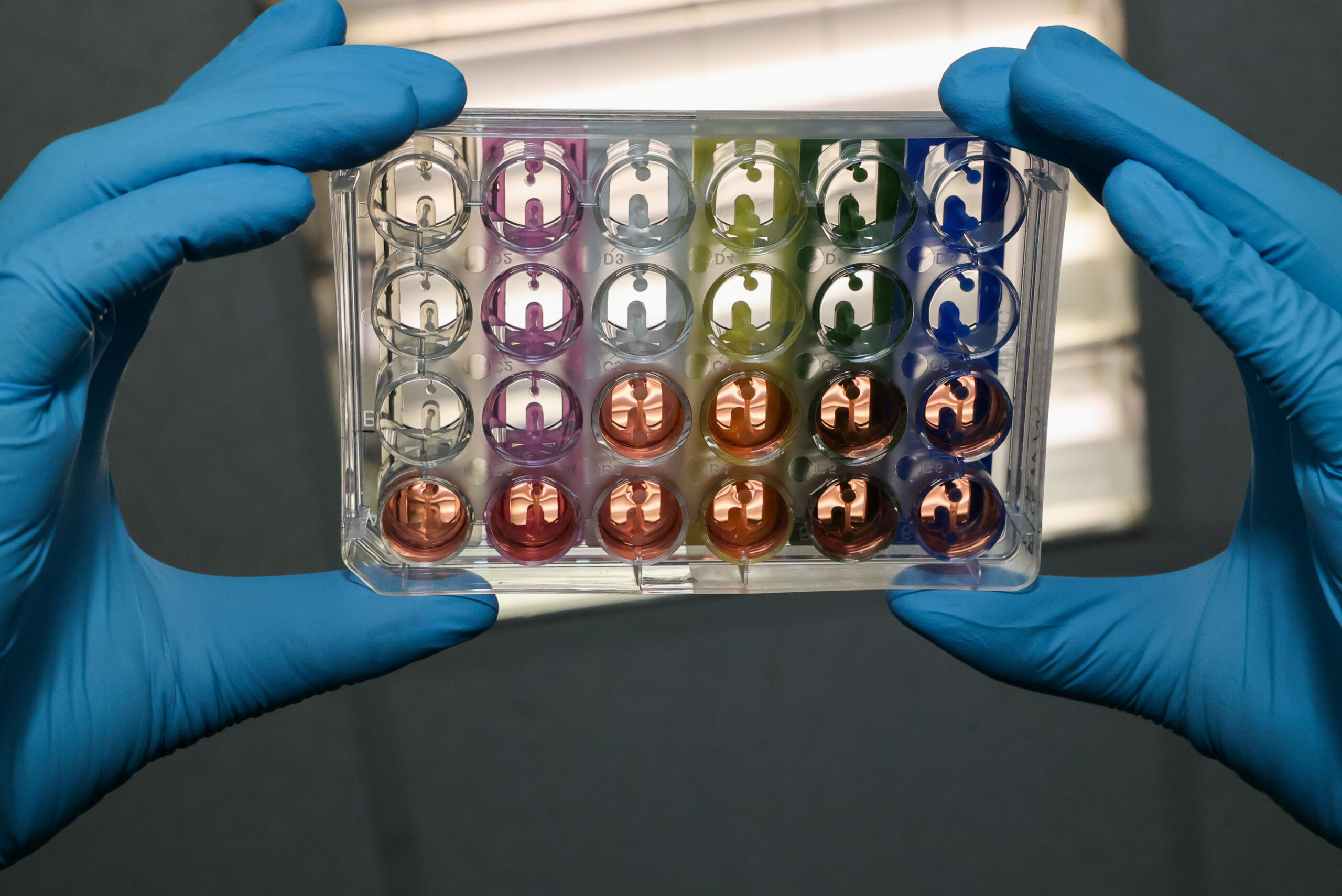
Phosphatidylserine improves the anti-inflammatory function of high density lipoproteins (HDL)


OUr founders


ISO 9001 Certified
OUr Institutionnal Supporters

Our network