
A team of researchers from the IHU ICAN has, for the first time, identified the role of epicardial adipose tissue (heart fat) and macrophages in the progression of atrial cardiomyopathy, which causes atrial fibrillation—the most common heart rhythm disorder worldwide.
Published on July 10, 2026, in *Circulation Research*, this scientific discovery broadens our understanding of the mechanisms underlying atrial fibrillation and opens up new avenues for developing treatments that target the early stages of the disease.
Understanding the Mechanisms That Prepare the Heart for Atrial Fibrillation
Atrial fibrillation is the most common heart rhythm disorder worldwide.
Its prevalence is rising rapidly due to an aging population, as well as the rise in cardiometabolic diseases, particularly obesity, high blood pressure, and diabetes.
Despite advances in treatment—particularly in ablating arrhythmogenic foci—this arrhythmia is most often recurrent and persistent.
Atrial fibrillation does not occur suddenly. It results from a gradual remodeling of the atria, known as atrial cardiomyopathy, characterized by changes in the electrical properties of the atria and fibrosis of the myocardial tissue.
Some figures
- 60 million people¹ worldwide live with atrial fibrillation (including 750,000 people in France),
- Its prevalence increases sharply with age and cardiometabolic diseases: 1 in 5 people are affected after age 45,
- Atrial fibrillation increases the risk of stroke by a factor of 52,
- Obesity, diabetes, and cardiometabolic diseases are among the leading modifiable risk factors.
Epicardial adipose tissue (the fat naturally present on the surface of the heart) is associated with an increased risk of atrial fibrillation.
However, its exact role in the progression of the disease is still poorly understood.
A First-of-Its-Kind Map of the Heart’s Immune Cells
A team of researchers from IHU ICAN, Sorbonne University, and Inserm, in collaboration with several French and international partners, has recently shed new light on the biological mechanisms that gradually lead to the remodeling of the heart’s atria.
The objective of this study was to identify the cellular mechanisms linking cardiac adipose tissue, inflammation, and atrial remodeling in order to better understand the early stages of the disease and identify new therapeutic targets.
To answer this question, the researchers drew on several of the most innovative technologies currently available in biomedical research:
- Spatial transcriptomics
- Single-cell RNA sequencing
- Multiphoton imaging
- CARS and SHG Microscopy
- Experimental and transgenic models of atrial cardiomyopathy
- Advanced Bioinformatics Analyses
1
Human cardiac tissue obtained from patients who had undergone surgery was first studied using spatial transcriptomics, a technology that allows for the visualization of gene expression directly within the tissue while preserving its anatomical organization.
2
These analyses were then combined with single-cell RNA sequencing, high-resolution multiphoton imaging, advanced microscopy techniques, and various experimental models that replicate the effects of obesity on the heart.
3
This integrated approach has made it possible to track, with unprecedented precision, the interactions between immune cells, adipose tissue, and cardiac muscle throughout the course of the disease.
A Major Discovery: Two Populations of Macrophages Orchestrate the Progression of the Disease
The study highlights a previously unknown mechanism: two distinct populations of macrophages are successively involved in atrial cardiomyopathy.
Lyve1+ macrophages, which are naturally present in cardiac adipose tissue, play a protective role. They accompany the expansion of epicardial adipose tissue and help maintain the organization of the heart muscle during the initial adaptations to metabolic stress.
As the disease progresses, this population decreases, while a second population—CCR2+ macrophages derived from blood cells—is recruited to areas where adipose tissue begins to be replaced by fibrosis. These inflammatory macrophages then promote atrial remodeling and dilation, creating conditions conducive to the development of atrial fibrillation.
The researchers also demonstrate that experimentally blocking this second population—CCR2+ macrophages—helps limit fibrosis and reduce the likelihood of developing episodes of atrial fibrillation.
A Paradigm Shift in the Understanding of Atrial Cardiomyopathy
Beyond identifying new cell populations, this study profoundly reshapes our understanding of the mechanisms underlying the disease.
Until now, epicardial adipose tissue has been viewed primarily as a factor associated with atrial fibrillation.
Researchers have shown that it is, in fact, a true biological actor, capable of locally orchestrating the inflammatory processes that gradually lead to the remodeling of the heart muscle.
“In other words, it’s not just the amount of fat that matters, but its biological activity and its interactions with the immune system.“
“This new perspective paves the way for the development of earlier biomarkers as well as new therapeutic strategies that directly target the mechanisms responsible for disease progression.“
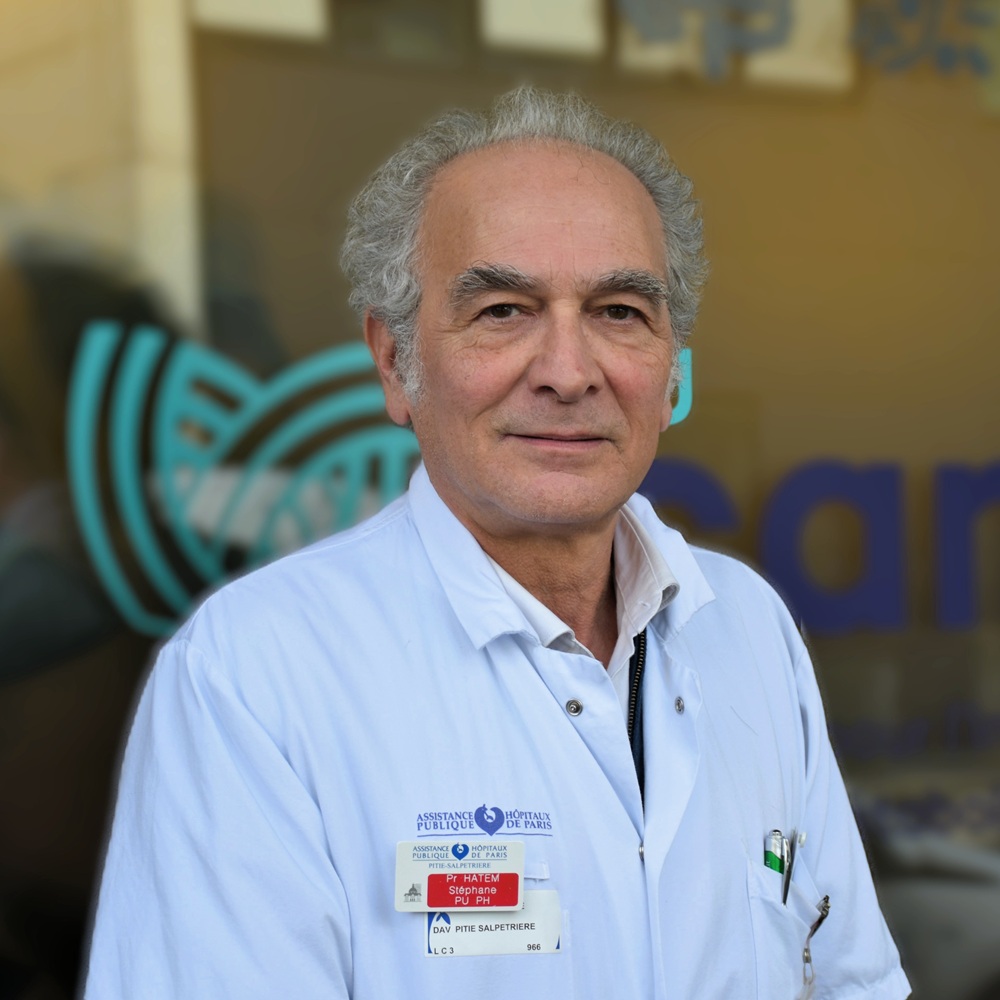
Prof. Stéphane Hatem
, Director of the IHU ICAN
A Breakthrough in Precision Medicine
By identifying previously unknown mechanisms underlying atrial cardiomyopathy, this study opens up significant possibilities for the future of medicine.
Ultimately, specifically targeting certain populations of macrophages could help slow down or even prevent atrial remodeling in patients at the highest risk for cardiometabolic diseases, even before atrial fibrillation develops.
This research could also help advance the development of new biomarkers that would allow for earlier identification of at-risk patients and the personalization of their care.
They fully demonstrate the potential of the translational research conducted at the IHU ICAN to transform basic scientific discoveries into innovations that benefit patients with cardiometabolic diseases.
“Understanding the very earliest stages of the disease is essential if we are ever to prevent its progression rather than treat its consequences. This study reveals a previously unknown biological mechanism linking cardiometabolic diseases, inflammation, and atrial fibrillation.”
“This research was made possible thanks to the expertise of the IHU ICAN, and it illustrates the importance of collaboration between basic and clinical research in accelerating the development of new therapeutic strategies that benefit patients.”
Prof. Stéphane Hatem
, Director of the IHU ICAN
The 3 Key Points to Remember
1. A New Map
immune cells present in cardiac adipose tissue, which can be used to develop new biomarkers for disease
progression
2. The discovery of a new biological mechanism
involving two populations of macrophages that succeed one another during the course of the disease, which may lead to the development of treatments that specifically target the macrophages involved in cardiac
remodeling
3. New Therapeutic Approaches
to intervene before irreversible damage occurs, and to identify at an earlier stage patients at risk for atrial fibrillation among those with cardiometabolic
diseases
An international collaboration
This study is the result of a collaboration between Sorbonne University, Inserm, IHU ICAN, the Pasteur Institute, the Indian Institute of Technology Delhi, and several clinical teams at Pitié-Salpêtrière Hospital.
It draws on the expertise developed at the IHU ICAN in cardiology, immunology, cell biology, transcriptomics, high-resolution imaging, bioinformatics, and translational research.
Among the co-authors from the IHU ICAN community are: L. Crepin, E. Trenquier, I. Abdou-Seini, A. de Raymond-Cahuzac, M. Ponnahia, N. Mougenot, S. Hatem, and N. Suffee.
The Role of the IHU ICAN
This study fully illustrates the scientific strategy of the IHU ICAN: to understand the fundamental mechanisms of cardiometabolic diseases in order to identify new therapeutic targets and accelerate their translation into clinical practice.
It draws on the institute’s complementary expertise in basic research, immunology, cardiology, cutting-edge imaging, bioinformatics, and clinical research, demonstrating the IHU ICAN’s ability to foster scientific innovations with high potential for translation into patient care.