Ultrasound, CT scans, MRI, PetScan… Doctors have a wide range of tools at their disposal to give them a better view of the disease. Once a simple aid to diagnosis, imaging is now an essential tool for improving diagnosis and managing treatment. Imaging is now capable of identifying abnormal mechanisms within tissues, i.e. the pathophysiological processes behind diseases that can be diagnosed earlier and more accurately. Finally, imaging is now a major interface between artificial intelligence and medicine.
AI in medical imaging
Artificial Intelligence (AI) is opening up new prospects for medical imaging , as it can take the analysis and interpretation of imaging further, thanks to its ability to integrate, combine and interpret a multitude of data in a very short space of time.
AI is capable of integrating data from the patient’s history, the history of their illness, the clinical context, biological parameters and data from other paraclinical examinations and previous examinations. The experience acquired through data integration, coupled with data from existing medical literature, enables AI to develop new diagnostic strategies, prognostic assessments and personalised patient management.
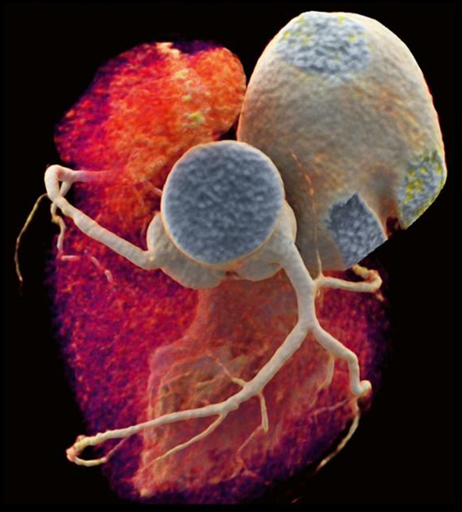
The new concept of imaging ‘biomarkers’ is now firmly established, and the advanced phenotyping made possible by techniques such as MRI, CT, ultrasound and optical imaging is opening up new avenues of exploration in humans, particularly in the field of cardiometabolic diseases.
The strengths of the ICAN IHU
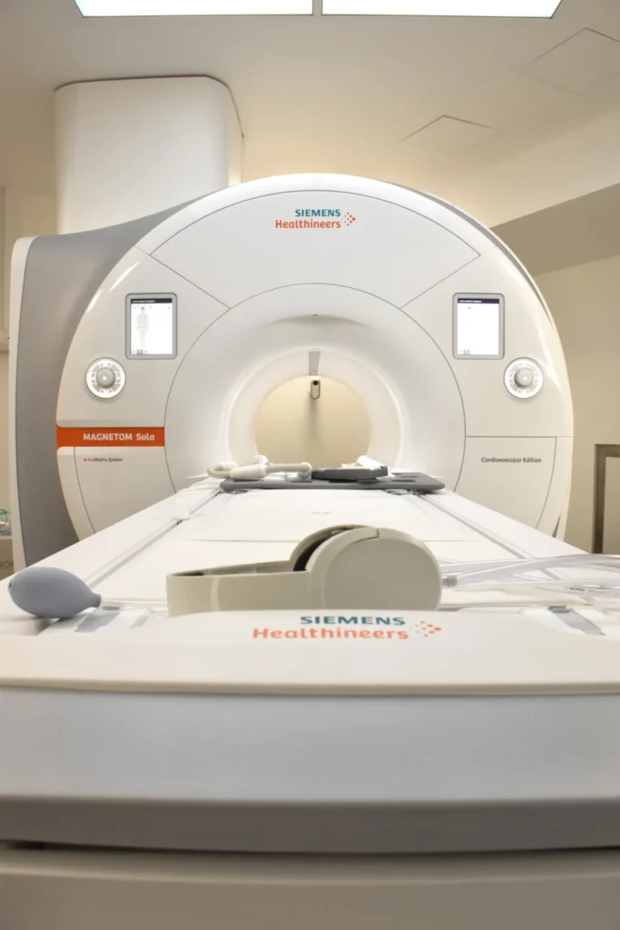
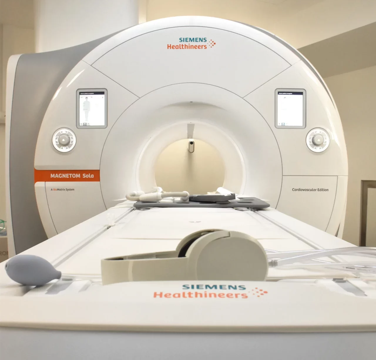
The ICAN IHU is positioning itself as a key player in the imaging of cardiovascular and metabolic diseases, thanks to its expert teams of researchers and clinicians and the establishment ofICAN Imaging, a cardiovascular and metabolic imaging platform that is unique in the Paris region, with the acquisition in 2020 of a latest-generation 1.5T cardiovascular MRI.